Back pain osteochondrosis
Back pain – osteochondrosis
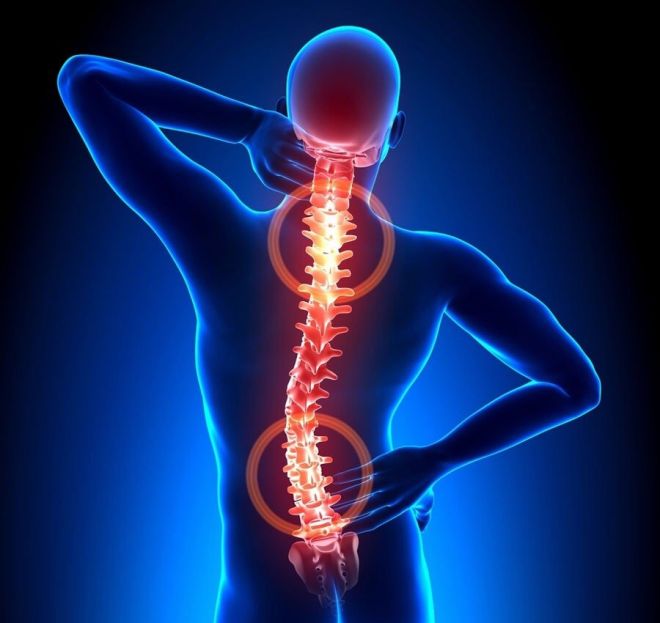
Osteochondrosis can be called a chronic disease in which the intervertebral discs and cartilage tissue are affected. The disease is widespread and affects people over forty years of age. The first symptoms can be found in twenty to thirty years. We can say that most back pain in people is caused by osteochondrosis. The human spine consists of several dozen vertebrae. To prevent friction of sensitive vertebrae with each other, so-called intervertebral discs are located between each pair. They perform the function of a damper in the spinal column. With osteochondrosis, blood circulation in the spinal column is disturbed, and in connection with this, a metabolic failure occurs. These processes also affect the intervertebral discs, when the intervertebral discs thin out during the disease and cease to perform their functions, then we can talk about the development of a disease called osteochondrosis of the spine. Osteochondrosis of the spine can develop in one part, for example, in the cervical, thoracic or lumbosacral, or affect the entire spine.
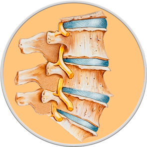
NORMAL SPINE
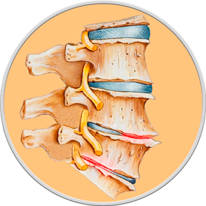
OSTEOCHONDROSIS
The diagnosis of osteochondrosis is made on the basis of the patient's complaints, objective examination data and X-ray examination. X-ray remains the simplest and most accessible method of examining joints to assess anatomical changes in the bone structure in the case of osteochondrosis. The method of X-ray diagnostics allows you to assess both the entire spinal column as a whole and its specific sections: cervical, thoracic and lumbosacral. X-ray examination allows you to determine the localization of the process, establish its nature and severity, and also identify the consequences of osteochondrosis, such as narrowing of the spinal canal, the presence of osteophytes, signs of spondylarthrosis. X-ray examination is performed, as a rule, in two projections: direct (in the supine position) and lateral or oblique projection. No special preparation for the examination is required, with the exception of x-ray examination of the lumbosacral region, before which bowel cleansing is required.
For a deeper assessment of the condition of the spinal column and its functional capabilities, it is necessary to perform an X-ray of the spine with functional tests, that is, while performing special exercises.
In the case of progression of osteochondrosis, pinched nerve roots, or suspected intervertebral hernias, magnetic resonance imaging is used for accurate diagnosis, which allows for the detection of changes in the soft tissues surrounding the spinal column.