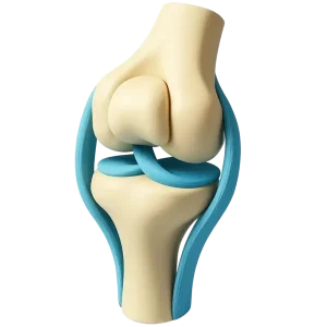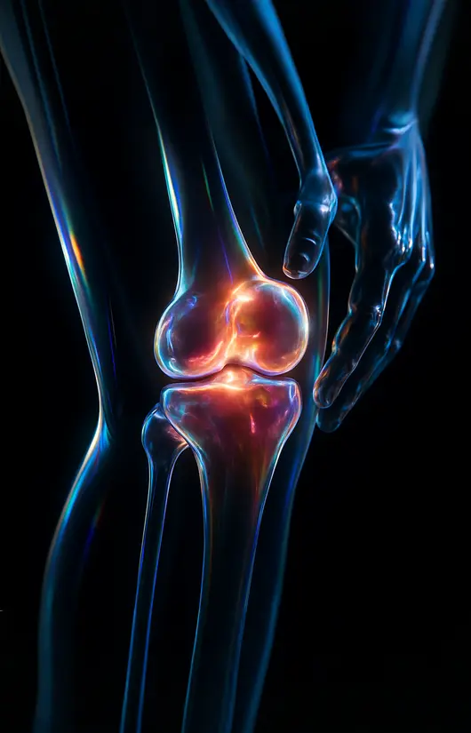
Симптоми при гонартрозі
Тривожні симптоми. У разі їх появи слід обов’язково звернутися до лікаря для проведення диференційного діагнозу.
- Morning stiffness for more than 30 minutes (possible rheumatic disease, collagenoses)
- Knee pain occurred after an infectious disease (possible infectious arthritis)
- Joint pain occurred after stress: hypothermia, surgery, etc. (possible rheumatic disease, collagenosis)
- Knee pain in patients with diabetes (consultation of an endocrinologist is required)
більше про симптоми гонартрозу >>
Risk factors
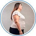 Excess body weightAccording to studies, obese women develop knee osteoarthritis 4 times more often than women of normal weight.
 Heredity. Joint diseases can be hereditary. In families where there have been cases of arthrosis, the likelihood of its occurrence is 2-3 times higher.
 Age. The older a person is, the higher the risk of the disease. This is due to a decrease in the ability of cartilage tissue to regenerate and a deterioration in metabolism in the joints.
 Increased loads. Hard physical labor and intense training can lead to microtrauma to joint tissues, which can lead to osteoarthritis.
Doctors' recommendations
- It is necessary to engage in swimming or other sports. At the same time, strictly
Dose the load on the joints. - Try to avoid injuries.
- Control your weight.
- Follow a diet that limits the consumption of fatty and spicy foods,
smoked meats, alcoholic beverages and other products that can negatively affect health. - If you are injured, you should immediately consult a doctor and complete a full course of treatment.
treatment. - After 35 years of age, you should periodically undergo preventive therapy courses. chondroprotectors.
- Try to drink enough water every day.
- Never self-medicate.