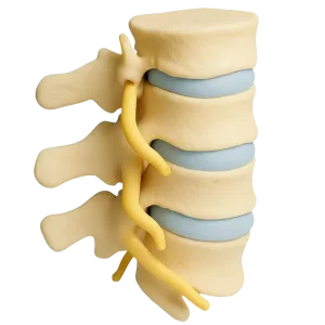
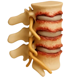
Symptoms of osteochondrosis

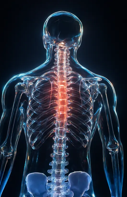
The main symptoms are back or neck pain, which worsens after physical exertion, prolonged sitting or sudden movements. Stiffness and decreased mobility of the affected area of the spine are often felt, especially in the morning. Headache, dizziness, tinnitus (in cervical osteochondrosis) or pain radiating to the limbs (in lumbar osteochondrosis) may occur. Tingling, numbness, a feeling of "pins and needles" in the arms or legs due to nerve compression often occurs. In severe cases, there is a violation of posture, decreased muscle strength, and limitation of limb functions. Symptoms progress gradually, so early diagnosis and treatment are important.

Depending on the location of the lesions, the symptoms of osteochondrosis may vary.

more about symptoms of osteochondrosis >>

Risk factors


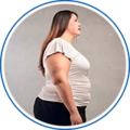
Excess body weightAccording to studies, obese women develop knee osteoarthritis 4 times more often than women of normal weight.

Heredity. Joint diseases can be hereditary. In families where there have been cases of arthrosis, the likelihood of its occurrence is 2-3 times higher.

 Age. The older a person is, the higher the risk of the disease. This is due to a decrease in the ability of cartilage tissue to regenerate and a deterioration in metabolism in the joints.


Increased loads. Hard physical labor and intense training can lead to microtrauma to joint tissues, which can lead to osteoarthritis.

Doctors' recommendations

  • It is necessary to engage in swimming or other sports. At the same time, strictly
    Dose the load on the joints.
  • Try to avoid injuries.
  • Control your weight.
  • Follow a diet that limits the consumption of fatty and spicy foods,
    smoked meats, alcoholic beverages and other products that can negatively affect health.
  • If you are injured, you should immediately consult a doctor and complete a full course of treatment.
    treatment.
  • After 35 years of age, you should periodically undergo preventive therapy courses. chondroprotectors.
  • Try to drink enough water every day.
  • Never self-medicate.
руйнування суглоба

Діагноз остеохондроз

Osteochondrosis can be called a chronic disease in which the intervertebral discs and cartilage tissue are affected. The disease is widespread and affects people over forty years of age. The first symptoms can be found in twenty to thirty years. We can say that most back pain in people is caused by osteochondrosis. The human spine consists of several dozen vertebrae. To prevent friction of sensitive vertebrae with each other, so-called intervertebral discs are located between each pair. They perform the function of a damper in the spinal column. With osteochondrosis, blood circulation in the spinal column is disturbed, and in connection with this, a metabolic failure occurs. These processes also affect the intervertebral discs, when the intervertebral discs thin out during the disease and cease to perform their functions, then we can talk about the development of a disease called osteochondrosis of the spine. Osteochondrosis of the spine can develop in one part, for example, in the cervical, thoracic or lumbosacral, or affect the entire spine.

Osteochondrosis
Osteochondrosis: Mechanism of spinal destruction

Healthy spine and affected discs

NORMAL SPINE
OSTEOCHONDROSIS
оцінка стану

Методи дослідження остеохондрозу

The diagnosis of osteochondrosis is made on the basis of the patient's complaints, objective examination data and X-ray examination. X-ray remains the simplest and most accessible method of examining joints to assess anatomical changes in the bone structure in the case of osteochondrosis. The method of X-ray diagnostics allows you to assess both the entire spinal column as a whole and its specific sections: cervical, thoracic and lumbosacral. X-ray examination allows you to determine the localization of the process, establish its nature and severity, and also identify the consequences of osteochondrosis, such as narrowing of the spinal canal, the presence of osteophytes, signs of spondylarthrosis. X-ray examination is performed, as a rule, in two projections: direct (in the supine position) and lateral or oblique projection. No special preparation for the examination is required, with the exception of x-ray examination of the lumbosacral region, before which bowel cleansing is required.

For a deeper assessment of the condition of the spinal column and its functional capabilities, it is necessary to perform an X-ray of the spine with functional tests, that is, while performing special exercises.

In the case of progression of osteochondrosis, pinched nerve roots, or suspected intervertebral hernias, magnetic resonance imaging is used for accurate diagnosis, which allows for the detection of changes in the soft tissues surrounding the spinal column.